TOMOGRAFÍA DENTAL 3D
Tomografía dental 3D en Costa Rica: planificación precisa para tratamientos complejos
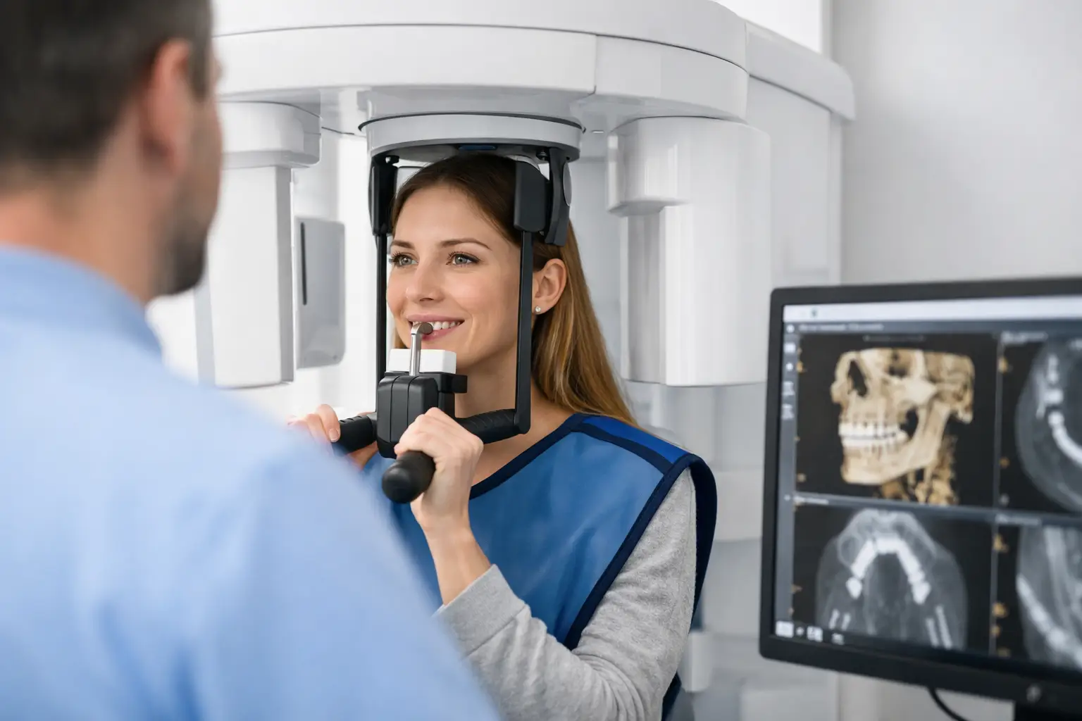
El sistema SCANORA® 3D de Sonrisa Para Todos ofrece imágenes tridimensionales de alta resolución con la menor dosis de radiación posible, siendo la herramienta esencial para la planificación de implantes, cirugías y tratamientos de alta complejidad.
- Tecnología SCANORA® 3D de última generación
- Imagen 3D en segundos con mínima radiación
- Indispensable para implantología y cirugía oral
- Archivo digital compartible con otros especialistas
Diagnóstico 3D de precisión con tecnología de clase mundial
Contact
Please provide us with your details and comments so we can connect you as soon as possible.
"*" indicates required fields
TOMOGRAFÍA DENTAL 3D
What is 3D dental tomography and why is it superior?
La tomografía computarizada de haz cónico (CBCT) es una técnica diagnóstica que obtiene imágenes tridimensionales de la estructura dental, maxilar y mandibular con una resolución sin precedentes. A diferencia de una radiografía convencional, que muestra una imagen plana en dos dimensiones, la CBCT permite visualizar cada estructura desde cualquier ángulo con exactitud milimétrica.
Ventajas sobre las radiografías convencionales:
- Imagen tridimensional vs. bidimensional plana
- Mayor resolución y detalle de estructuras óseas
- Visualización de nervios, senos y tejidos blandos
- Permite medir distancias y ángulos con precisión
- Esencial para implantología guiada y cirugía oral
APLICACIONES CLÍNICAS
¿Para qué tratamientos se utiliza la tomografía 3D?
La CBCT es la herramienta de diagnóstico de referencia en múltiples especialidades odontológicas.
MÁS RECOMENDADO
Implantología
Planificación guiada de implantes dentales
- Medición exacta del volumen óseo
- Localización precisa del nervio dentario
- Posicionamiento virtual del implante
- Reducción de riesgos en cirugía
Cirugía oral
Cordales, quistes y casos complejos
- Localización de dientes incluidos
- Evaluación de quistes y patologías óseas
- Planificación de extracciones complejas
- Análisis de tejidos adyacentes
Endodoncia y ortodoncia
Anatomía radicular y análisis esquelético
- Detecta conductos adicionales no visibles
- Análisis 3D de posición dental y esquelética
- Evaluación de reabsorción radicular
- Planificación de movimientos complejos

TECNOLOGÍA SCANORA® 3D
El equipo más avanzado en diagnóstico dental tridimensional
Características técnicas:
- Sistema SCANORA® 3D de Soredex®
- Múltiples campos de visión (FOV) seleccionables
- Alta resolución con dosis de radiación optimizada
- Reconstrucción 3D inmediata en pantalla
Ventajas para el paciente:
- Procedimiento rápido: menos de 15 segundos de exposición
- Posición cómoda durante la toma
- Sin molestias ni preparación especial
- Resultados disponibles de inmediato
¿CÓMO ES EL PROCESO?
Realizarse una tomografía dental 3D es rápido y sin complicaciones
Todo el proceso toma menos de 30 minutos desde que llegas a la clínica.
1
Posicionamiento del paciente
Te ubicamos correctamente en el equipo SCANORA® 3D para garantizar una imagen de calidad en el área de interés clínico.
2
Toma de la imagen
La exposición dura menos de 15 segundos. El equipo gira alrededor de la cabeza capturando cientos de imágenes que se reconstruyen en 3D.
3
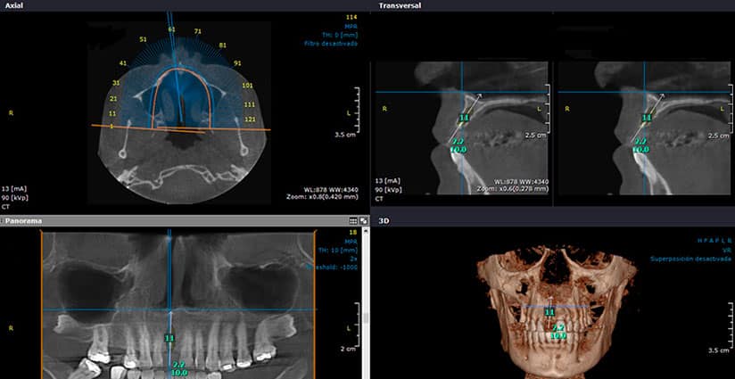
Reconstrucción y análisis
El software genera de inmediato la imagen tridimensional, que el especialista analiza en distintos planos y ángulos.
4
Treatment plan
Con base en la tomografía, el especialista diseña el plan quirúrgico o terapéutico personalizado para tu caso.
SOFTWARE DIAGNÓSTICO
Análisis digital 3D para diagnósticos de alta precisión
El sistema SCANORA® 3D trabaja con software de última generación que permite visualizar la imagen desde cualquier corte, medir estructuras con precisión milimétrica, planificar la posición ideal de los implantes de forma virtual y exportar los datos a guías quirúrgicas. Todo esto se traduce en tratamientos más seguros, predecibles y eficientes.
- Medición de densidad y volumen óseo
- Planificación virtual de implantes en 3D
- Exportación a guías quirúrgicas
- Análisis de nervios y estructuras anatómicas

¿QUIÉN LA NECESITA?
Casos que requieren tomografía dental 3D
No todos los casos requieren tomografía 3D, pero en los siguientes escenarios es prácticamente indispensable:
- Planificación de implantes dentales unitarios o múltiples
- Evaluación de cordales incluidas o impactadas
- Tratamientos de endodoncia en anatomía compleja
- Diagnóstico de quistes, tumores o patologías óseas
- Planificación de cirugía ortognática
- Casos de ortodoncia con dientes retenidos
POR QUÉ ELEGIRNOS
Diagnóstico 3D integrado con tu plan de tratamiento
SCANORA® 3D
Contamos con el sistema SCANORA® de Soredex®, uno de los equipos de tomografía dental más reconocidos por su calidad de imagen y seguridad radiológica.
Planificación quirúrgica digital
Nuestros especialistas utilizan el análisis 3D para diseñar guías quirúrgicas y planificar cada caso con precisión milimétrica.
25+ años de experiencia
Décadas de experiencia en diagnóstico e interpretación de imágenes dentales para casos de implantología, cirugía y ortodoncia.
Expediente digital completo
Todas las tomografías se archivan digitalmente en tu expediente y están disponibles para consulta en cualquier momento.
TESTIMONIOS
Pacientes que se beneficiaron de nuestro diagnóstico 3D
★★★★★
“La tomografía 3D les permitió planificar mis implantes de forma exacta. El resultado fue perfecto y sin complicaciones en la cirugía.”
Luis F.
San José, Costa Rica
★★★★★
“Vine desde Florida para colocarme implantes. La tomografía previa fue fundamental — gracias a ella detectaron una condición que otro dentista no había visto.”
Sandra M.
Miami, Florida
★★★★★
“Me tranquilizó mucho saber que todo estaba planificado al detalle antes de entrar a cirugía. La tomografía 3D hizo toda la diferencia.”
Carmen V.
Heredia, Costa Rica

INVERSIÓN EN TU SALUD
Tomografía dental 3D: diagnóstico completo a precio accesible
El costo de una tomografía dental 3D en Costa Rica es hasta un 70% menor que en Estados Unidos, con exactamente la misma tecnología. Como parte de tu plan de tratamiento integral, puede estar incluida sin costo adicional según el caso.
- Incluida en la planificación de implantes dentales
- Precio significativamente menor que en EE.UU.
- Resultados disponibles de inmediato
- Análisis e interpretación por especialista incluida
Muchos pacientes internacionales inician su tratamiento con una tomografía 3D en Costa Rica, lo que les permite obtener un diagnóstico completo y definitivo a una fracción del costo en su país.
Financing available

PREGUNTAS FRECUENTES
Todo lo que debes saber sobre la tomografía dental 3D
¿En qué se diferencia la tomografía 3D de una radiografía panorámica?
La radiografía panorámica es una imagen bidimensional que muestra toda la dentición en un plano. La tomografía 3D genera una imagen tridimensional con cortes en todos los planos, permitiendo medir estructuras, evaluar densidad ósea y planificar implantes con precisión milimétrica.
¿Es segura la tomografía dental 3D?
Sí. La tomografía CBCT utiliza dosis de radiación considerablemente menores que una tomografía médica convencional, y significativamente inferiores a los límites de seguridad establecidos. El equipo SCANORA® 3D está diseñado para optimizar la dosis sin comprometer la calidad de imagen.
¿Cuánto tiempo dura el proceso?
La toma de la imagen dura menos de 15 segundos. El proceso completo, incluyendo posicionamiento y análisis, toma aproximadamente 15 a 30 minutos dependiendo de la complejidad del caso.
¿Debo prepararme de alguna forma antes de la tomografía?
No se requiere preparación especial. Solo se recomienda retirar objetos metálicos de la cabeza y el cuello (aretes, cadenas, armazones metálicos) antes de la toma para evitar artefactos en la imagen.
¿Puedo solicitar una copia de mi tomografía?
Sí. La tomografía se archiva en formato digital en su expediente. Puede solicitarla en cualquier momento para compartir con otro especialista. Se entrega en formato estándar compatible con la mayoría de los softwares de visualización.
¿Es necesaria la tomografía para todos los implantes?
Sí, es parte esencial del protocolo de planificación. Nos permite medir el volumen y la densidad ósea, identificar la posición exacta del nervio dentario y diseñar la cirugía con mayor seguridad. Sin tomografía, no podemos garantizar un resultado predecible.
¿Necesitas una tomografía dental 3D para planificar tu tratamiento?
Con el sistema SCANORA® 3D obtenemos el diagnóstico más completo disponible. Agenda tu consulta y recibe un análisis detallado de tu caso.